Jho Institute for Minimally Invasive Neurosurgery Department of Neuroendoscopy
Spine Diseases
Brain Diseases
Hemifacial Spasm Surgery: Dr. Jho's Endoscopic Microvascular Decompression Surgery for Hemifacial Spasm
Dr. Jho's Minimally Invasive Endoscopic Microvascular Decompression Surgery for Hemifacial Spasm
Professor & Chair, Department of Neuroendoscopy
Jho Institute for Minimally Invasive Neurosurgery
Having worked for approximately 20 years with Professor Jannetta (who pioneered the development of various Jannetta procedures for microvascular decompression), Dr. Jho has developed minimally invasive endoscopic microvascular decompression surgery for hemifacial spasm and other cranial nerve disorders. Microvascular decompression surgery was originally pioneered by Professor Jannetta, who has spent his entire neurosurgery career exploring various neurovascular compression syndromes.
Microvascular decompression (which consists of placement of small synthetic sponges between the compressing blood vessels and the affected cranial nerves) carries a good chance of relieving cranial nerve compression symptoms such as hemifacial spasm.
Cranial nerve surgery is done through a small skull opening behind the ear and is referred to as retromastoid craniectomy. Skin incisions are usually two inches in length. Surgery is performed under the endoscopic visualization.
When blood vessels cross and compress cranial nerves, various characteristic symptoms develop depending upon which cranial nerves are compressed. Trigeminal neuralgia develops by blood vessel compression of the trigeminal nerve, hemifacial spasm by compression of the facial nerve, intractable positional vertigo by compression of the vestibular nerve, tinnitus by compression of the cochlear nerve, glossopharyngeal neuralgia by compression on the glossopharyngeal nerve, and spasmodic torticollis by pressure on the spinal accessory nerve and upper cervical nerves.
A: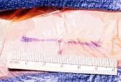 B:
B: 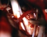
B: Figure 1. For microvascular decompression, a 4-cm skin incision is made behind the patient's ear (A). An intraoperative photograph displays arterial compression of the facial nerve in a patient with hemifacial spasm (B).
Practice Manager: Robin A. Coret
Tel : (412) 359-6110
Fax : (412) 359-8339
Address : JHO Institute for Minimally Invasive Neurosurgery
Department of Neuroendoscopy
Sixth Floor, South Tower
Allegheny General Hospital
320 East North Avenue
Pittsburgh, PA 15212-4772


Copyright 2002-2032