Jho Institute for Minimally Invasive Neurosurgery Department of Neuroendoscopy
Spine Diseases
Brain Diseases
Spinal Cord Tumor Surgery: Dr. Jho's Anterolateral or Posterolateral Approach for Spinal Cord Tumors
Dr. Jho's Minimally Invasive Spinal Cord Tumor Surgery: Anterolateral or Posterolateral Approach for Spinal Cord Tumors, Endoscopic Spinal Cord Tumor Surgery
Professor & Chair, Department of Neuroendoscopy
Jho Institute for Minimally Invasive Neurosurgery
Under a minimally invasive surgical strategy in spine surgery, Dr. Jho has developed various spinal tumor surgeries such as an anterolateral approach and a posterolateral approach for cervical spine tumors, and minimally invasive endoscopic approaches for cervical, thoracic and lumbar spine tumors. These minimally invasive approaches accomplish adequate tumor removal with maximal protection of the spinal cord and spinal nerves. These approaches often eliminate the need for bone fusion also.
Spinal cord tumors include spinal bone tumors, tumors located in the spinal canal but outside the spinal cord itself (extramedullary tumors), and tumors of the spinal cord itself (intramedullary).
Spinal bone tumors can be divided into cancerous tumors and benign tumors. Cancerous spinal bone tumors include metastatic cancers and primary bone cancers. Although any type of cancers can spread to the spinal bone, the most common ones include lung cancers, breast cancers, prostate cancers, kidney cancers, and lymphomas. Primary cancerous spinal bone tumors are relatively uncommon and include chordomas, osteosarcomas, chondrosarcomas, Ewing's sarcomas, multiple myelomas, lymphomas etc. Treatments for these malignant tumors must involve multimodality measures including surgery, chemotherapy and radiation. Benign spinal bone tumors include osteoid osteomas, osteoblastomas, osteochondromas, giant cell tumors, aneurysmal bone cysts, plasmacytomas, hemangiomas, etc. Treatments for these benign tumors have to be individualized depending on the type of pathology and their symptoms.
Two common extramedullary tumors are neurofibromas and meningiomas. Neurofibromas can occur as a sporadic condition or as a genetic disorder such as neurofibromatosis. These are benign tumors that often require surgical removal.
Although hemangioblastomas, epidermoid, dermoid, and others can rarely occur, the two most common intramedullary tumors are astrocytomas and ependymomas. Treatment involves primarily surgical resection. Radiation treatments may be required in some cases as an adjunctive treatment.
Anterolateral Approach to Spinal Cord Tumors
A: 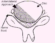 B:
B: 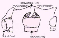
 B:
B: 
Figure 1. Schematic drawings, axial (A) and lateral oblique view (B) demonstrate exposure of a spinal cord tumor through a small hole of anterior microforaminotomy (anterolateral approach).
A: 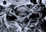 B:
B: 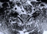
 B:
B: 
Figure 2. MR scans, axial views, show a spinal cord meningioma (A, an oval gray shadow at the center) preoperatively, and complete removal of the tumor postoperatively (B). Surgical access tract is demonstrated in a white shadow by contrast enhancement.
A: 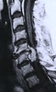 B:
B: 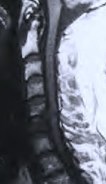
 B:
B: 
Figure 3. MR scans, sagittal views, reveal a spinal cord tumor and bony spurs at the C5-6 level preoperatively (A, a white shadow) and total removal of the tumor and bony spurs postoperatively (B). In this technique, bone graft fusion and metal implants are not necessary. Patients are hospitalized only overnight and do not need cervical collars postoperatively.
A: 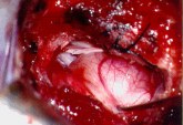 B:
B: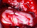
 B:
B:
Figure 4. These intraoperative photographs were taken during an anterolateral approach to a spinal cord tumor. A neurofibroma is exposed through an anterolateral microforaminotomy hole. The upper (A) and lower (B) edge of the tumor are demonstrated.
Posterolateral Approach for Spinal Cord Tumors
A: 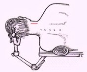 B:
B: 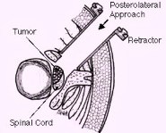
 B:
B: 
Figure 1. Schematic drawings show a patient's positioning, a line of a skin incision (3-5 cm) (A), and a surgical approach (B).
A: 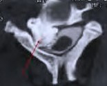 B:
B: 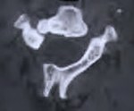
 B:
B: 
Figure 2. A bony tumor (osteoma) is demonstrated in a postmyelographic CT scan preoperatively (A, arrow), and total removal of the tumor is confirmed in a postoperative CT scan (B).
A: 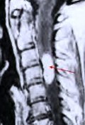 B:
B: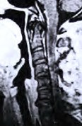 C:
C: 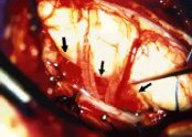
 B:
B: C:
C: 
Figure 3. A preoperative MR scan, sagittal view, reveals a spinal cord tumor (hemangioblastoma) (A, arrow). A postoperative MR scan, sagittal view, confirms total removal of the tumor (B). An intraoperative photograph demonstrates the lateral aspect of the stretched spinal cord and the tumor (C, arrows).
Facts About These Surgeries
Discussion
Both of these approaches, the anterolateral approach (which means the surgeon approaches from the front and to the side), and the posterolateral approach (which indicates the surgeon is working from the back and to the side), are bone-sparing procedures which focus on maintaining the structural stability of the spine. Conventional procedures often require extensive bone removal from the spine which then necessitates fusion and/or placement of metal implants to provide stability. The anterolateral approach spares bone by using small anterior foraminotomy holes (made for tumor access) and allows preservation of the structural integrity of the bone. The posterolateral approach spares bone by utilizing a limited hemilaminectomy. Most patients are able to be discharged the day following surgery. Various minimally invasive endoscopic techniques have been also used for different types of spinal cord tumors.
Definitions
foraminotomy - enlargement of an opening or passageway for a nerve root.
hemilaminectomy - excision of a vertebral lamina on one side.
References
Practice Manager: Robin A. Coret
Tel : (412) 359-6110
Fax : (412) 359-8339
Address : JHO Institute for Minimally Invasive Neurosurgery
Department of Neuroendoscopy
Sixth Floor, South Tower
Allegheny General Hospital
320 East North Avenue
Pittsburgh, PA 15212-4772
Copyright 2002-2032
